We are switching to a paperless form system to make your visit faster and easier. All patients will need to update their forms. Click here to learn more
A cataract is a loss of transparency, or clouding, of the eye lens. The eye lens plays a vital role in focusing images on the retina. A cloudy lens interferes with light passing through to the retina, the light-sensing layer of cells at the back of the eye. Compare a cataract to looking at the world through a foggy or blurry window. Light rays do not focus clearly if the lens loses its clarity, as it does when a cataract develops. Glasses or contact lenses cannot sharpen vision if a cataract is present.

The most common cause of a cataract is aging. As you get older, chemical changes in your lens make it less transparent. The loss of transparency may be so mild that vision is hardly affected or so severe that no shapes or movements are seen, only light and dark. You have a cataract when the lens gets cloudy enough to obstruct vision to any significant degree.
Other causes of cataracts include trauma, medications like steroids, systemic diseases such as diabetes, and prolonged exposure to ultraviolet light. Occasionally, babies are born with a cataract. Glasses or contact lenses cannot sharpen your vision if a cataract is present.
Reducing the amount of ultraviolet light exposure by wearing a wide-brim hat and sunglasses may reduce your risk for developing a cataract, but once set, there is no cure except to have the cataract surgically removed. Outpatient surgical procedures remove the cataract either through a small incision (phacoemulsification) or a large incision (extracapsular extraction). The time to have the surgical procedure is when your vision is bad enough to interfere with your lifestyle.
Cataract formation is a slow, progressive, and painless decrease in vision. Ironically as the lens gets harder, farsighted (hyperopic) people experience improved distance vision and are less dependent on glasses. However, nearsighted (myopic) people become more nearsighted, causing distance vision to worsen.
Request an appointmentA cataract is detected during our comprehensive eye exams. Your eyes will be dilated, so the pupils are wide open, enabling our medical team to look for signs of a cataract with a slit lamp, along with checking your retina and optic nerve. We will also do a refraction to test your visual acuity.
The treatment for a cataract is to remove the lens and implant an Intraocular Lens (IOL). An IOL is a tiny, lightweight, clear plastic disk placed in the eye during cataract surgery. Intraocular lenses have many advantages. The IOL remains in the eye after surgery, unlike contact lenses, which must be removed, cleaned, and reinserted. An IOL replaces the focusing power of the eye's natural lens. The rapid evolution of IOL designs, materials, and implant techniques has made them a safe and practical way to restore normal vision after cataract surgery.
One and a half million people have a cataract procedure every year, and 95% achieve success. As with any surgery, complications may occur during or after, and some are severe enough to limit vision. In most cases, vision, as well as the quality of life, improves.
Laser-assisted cataract surgery is performed as an outpatient procedure, typically under a local anesthetic (using eye drops) or sometimes with general anesthesia. The patient feels no pain, but you may need to avoid bending over or lifting heavy objects during recovery. It can take up to six weeks to achieve the best-corrected vision.
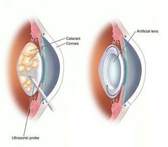
Phacoemulsification is a surgical method in which an ultrasonic oscillating probe is inserted into the eye. The probe breaks up the center of the lens. The fragments are suctioned from the eye at the same time.
A cataract is removed in tiny pieces with a small incision is made that does not require sutures to close. Most of the lens capsule is left behind, and a foldable intraocular lens implant, or IOL, is placed permanently inside to help focus light onto the retina. Vision returns quickly, and one can resume normal activities within a short period.
During cataract surgery, your eye surgeon will remove your eye’s cloudy natural lens. Then he or she will replace it with an artificial lens. This new lens is called an intraocular lens (or IOL). When you decide to have cataract surgery, your doctor will talk with you about IOLs and how they work.
People who have had cataract surgery may have their vision become hazy again years later. This is usually because the lens capsule has become cloudy. The capsule is the part of your eye that holds the IOL in place. Your ophthalmologist can use a laser to open the cloudy capsule and restore clear vision. This is called a capsulotomy.
Cataracts are a very common reason people lose vision, but they can be treated. You and your ophthalmologist should discuss your cataract symptoms. Together you can decide whether you are ready for cataract surgery.
The cataract surgeons at the Geneva Eye Clinic are board-certified and specialize in the diagnosis, assessment and surgical treatment of your cataracts. Geneva Eye Clinic offers you the latest in technology, especially when it comes to the diagnosis and treatment of cataracts.
Cataract Surgery Specialists at Geneva Eye Clinic
There are many types of IOLs, each with its own pros and cons. Below are some general categories. Ask your ophthalmologist about which type of IOL is best for you.
Monofocal: This is the type of IOL that most people select. Monofocal lenses have one focusing power. This means they sharpen either your distance, mid-range or close-up vision.
Toric: This lens is considered if you have a significant amount of astigmatism (an irregularly shaped cornea). These lenses improve how light hits your retina, allowing you to have a sharper, clearer vision. Toric lenses are available in monofocal, multifocal, extended depth of focus (EDOF) or accommodative models.
Multifocal: This lens makes it possible for patients to see near, intermediate, and distance without eyeglasses. Multifocal lenses contain several focal zones. Your brain adjusts to these zones and chooses the focusing power you need for any given task (like driving or reading).
Extended Depth of Focus (EDOF): This lens creates a single elongated focal point and provides an uninterrupted range of vision. These lenses give you excellent distance vision along with improvements in your mid-range vision (for tasks such as computer use). You may still need to use glasses for close-up tasks like reading.
Light Adjustable Lens: This is the first and only lens that can be customized after cataract surgery. A series of UV light treatment procedures, customize your lens prescription to bring you as close to your desired visual outcome as possible. This is still a type of monofocal lens, so glasses will be necessary for reading or driving.
Traditional Cataract Surgery
Cataract Surgery with Astigmatism Correction
Cataract Surgery for Distance Intermediate and Near Vision
Cataract Surgery with Customized Vision
Intraocular Lens |
|||||
|---|---|---|---|---|---|
| Monofocal (Standard) | Toric (Astigmatism) | Extended Depth of Focus (EDOF) | Multifocal | Light Adjustable Lens | |
| Customizable After Surgery | No | No | No | No | Yes |
| Astigmatism Correction | No | Yes | Yes | Yes | Yes |
| Zones of clear vision without glasses | Glasses likely needed at all distances for best vision | Distance | Distance, Intermediate | Distance, Intermediate, Near | Distance, Intermediate, Variable, Near (blended vision) |
| Strengths | Most affordable | Excellent distance vision without glasses | Excellent distance and intermediate vision | Excellent full range of vision (distance, intermediate, & near) | Allows unparalleled precision as the power can be adjusted after surgery |
| Weaknesses | Glasses likely needed at all distances for best vision | Glasses required for intermediate and near tasks | May need glasses for near tasks and low light (within 20 inches) | Low risk of glare and halos given lens design, including low risk of glare | Requires UV protective glasses for several weeks after surgery and in office adjustments to perfect the outcome. May need glasses for fine print. |
| Optimal patient | Doesn't mind wearing glasses or can't afford other options | Prioritizes distance activities, ok with glasses for computer and reading | Fits with modern lifestyle, ok with glasses for reading and low light | Needs full range of vision, ok with low risk of bothersome halos | Can afford truly customizable vision, prior refractive surgery patients |
| Good choice with coexistent ocular disease | Yes | Yes | Probably | No | Yes |
Find information on Cataracts in children in Pediatric Eye Care.