We are switching to a paperless form system to make your visit faster and easier. All patients will need to update their forms. Click here to learn more
To obtain your best-corrected vision, no matter your needs, the medical team at Geneva Eye Clinic performs many different tests. A technician will complete the first part of your comprehensive exam, including tests for visual acuity, eye pressure, and refraction. Next, the Ophthalmologist will test for common eye diseases and assess how your eyes work together. Follow-up appointments for recommended treatment may be necessary.
Your pupils will be dilated (widen your pupils) with eye drops to view the back of your eyes, the retina, and optic nerve. The dilating drops take approximately 30 minutes to take full effect. Your eyes may remain dilated for 4 to 6 hours. You will be sensitive to bright light and possibly blurry for near vision. We have disposable sunglasses available for your comfort. You may wish to have a driver with you, especially if this is the first time your eyes will be dilated.
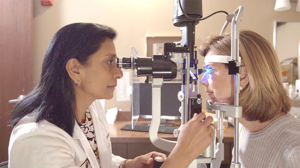
The doctor will perform the rest of your exam after your eyes are fully dilated. This usually entails using a microscope called a slit lamp to view the front structures of your eyes and bright lights to view the retina and optic nerves. While potentially uncomfortable, it will not harm your eyes. The doctor will discuss the exam findings with you and any treatment plans if needed.
A comprehensive eye exam takes approximately two hours. Please anticipate your visit to require this length of time.
Part of every eye exam, the eye chart test allows the doctor to measure your ability to see at varying distances.
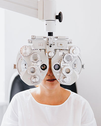
A refraction checks the prescription of your eyes and can be used to obtain glasses. A refraction test might be included even if you do not wish to get new glasses. Performing refraction shows the doctor your best-corrected vision and the best vision you can attain. A decrease in visual acuity can be the first sign that something is wrong. Knowing your best-corrected vision can help the doctor monitor changes in eye conditions or help to decide when cataract surgery is necessary. For children, especially preverbal children, the eye doctor will use additional expertise to perform this specialized measurement, whether or not the child ultimately requires glasses.
This instrument measures pressure within the eye: A high-pressure reading could be a sign of glaucoma. Before the test, numbing drops are placed in the eyes. The numbing drops do not affect your vision. In most cases, it is not necessary to measure eye pressure in children.
The slit lamp is used primarily to view the eye's anterior (front) structures, such as the iris, lens, and cornea. It is a microscope with a light attached, allowing examination of the eye under high magnification. Special lenses also examine the vitreous and the back of the eye.
The fundus exam consists of using a bright light to visualize the back structures of the eye, the retina, and optic nerve, through a dilated pupil. The eye is the only body part to see blood vessels without surgery. The doctor will check the health of your eyes and look for changes consistent with diabetes, macular degeneration, glaucoma, or other diseases.
A consultation exam is more extensive than a comprehensive eye exam and typically lasts several hours. Usually, another physician has referred you to diagnose and treat a specific condition that they don't specialize in or treat themselves. An in-depth health history, including family members, will be obtained to aid in the diagnosis. Additional special testing may be performed in the office, and labs or x-rays may be ordered. Communication with your primary care doctor or referring doctor may be necessary to coordinate your care efficiently.
These tests are only performed when indicated.
This test enables the doctor to determine if there is a loss of peripheral (side) vision, indicating glaucoma or neuro-ophthalmic conditions.
OCT is a crucial tool for diagnosing and managing conditions such as macular degeneration, diabetic retinopathy, retinal vein occlusion, and glaucoma. This test uses a beam of light to view the layers of the retina.
It is used in ophthalmology as a form of documentation the doctor uses to compare your eye at the current exam versus previous exams. This is helpful with glaucoma to monitor the size and shape of the optic nerves. It is also beneficial to look for any changes in a nevus (freckle in the retina) that could be significant over time.
Fluorescein Angiography helps your doctor see what is happening in the circulatory system of your retina, highlighting any abnormalities that may be present. A dye is injected into a vein in your arm. As the dye enters the blood vessels, photographs are taken of the back of your eye. These photos will help the doctor see many things, such as abnormal blood vessels and inadequate circulation areas. This procedure is performed in the office. Your eyes will be dilated. We recommend having someone drive you home from your appointment.
Before being assessed for glasses or contact lenses, you will need to have a comprehensive eye examination to ensure that your eyes are healthy.
Careful measurements, along with refraction, assist the doctor in deciding your prescription. Please bring glasses, reading glasses, or contacts to your appointment if you currently wear them.
During a low vision evaluation, a patient is assessed for aids to enhance remaining vision after a loss due to such conditions as macular degeneration, glaucoma, or other ocular diseases. When conventional glasses do not meet the patient's visual needs, various optical and non-optical devices are assessed, ranging from simple optical magnifiers to telescopic glasses or electronic magnifiers. Even though low vision aids do not replace lost vision, they can improve independence and your quality of life. It is essential for patients coming in for a low vision exam to bring all their glasses and vision devices.
Before being fit with contact lenses, you will need to have a comprehensive eye examination to ensure that your eyes are healthy. Careful measurements assist the doctor in deciding which lenses may be right for you. If you wear contacts lenses, please wear them to your appointment, bring your glasses and any information about your contacts such as the prescription or boxes.
Contact Lens Fitting Exam is for the first-time contact lens wearer. This exam will take more time due to instruction and training on insertion and removal of the lenses and reviewing lens care systems.
Contact Lens Refit Exam is for current contact lens wearers that may want or need another type of contact lens. Several lenses may be fit at this exam, and training is rarely required. Specialty contact lenses for astigmatism and keratoconus are available, along with multifocal contact lenses.